 |
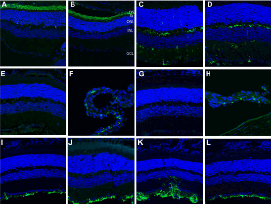
| Figure 7: Immunolocalization of resident retinal proteins and inflammatory markers in subretinal polymer injected eyes and control 1 week post-injection. Rhodopsin immunolabeling shows no difference in localization of rhodopsin in (A) control non-injected and (B) subretinal biopolymer injected eyes. Iba1-positive microglial cells are localized to the ganglion cell layer, inner plexiform layer and outer plexiform layer both (C) control subretinal BSS-injected and (D) subretinal biopolymer-injected eyes. F4/80 immunoreactivity was not observed in either (E) control BSS-injected or (G) subretinal biopolymer eyes. As positive controls, the ciliary body from the same section has F4/80-positive macrophages in both (F) control BSS-injected and (H) subretinal biopolymer-injected eyes. GFAP labeling in (I) non-injected control eye, (J) BSS-injected control eye and (K) subretinal biopolymer-injected eye at the site of needle entry and (L) subretinal biopolymer-injected eye distal to the injection site. Moderate proliferation of GFAP-positive Müller cells is seen at the injection site but not distal to the injection site in subretinal biopolymer eyes. |