|
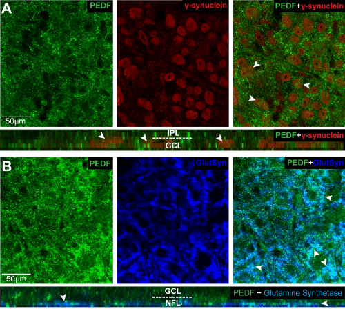
| Figure 2: PEDF is associated with RGCs and Müller cell endfeet in the ganglion cell and nerve fiber layers. A. Representative confocal micrographs with orthogonal view (bottom panel) of wholemount retina from naïve mice co-immunolabeled with antibodies against PEDF (green) and the RGC marker γ-synuclein (red). PEDF localizes to the area surrounding PEDF γ-synuclein+ RGCs in the ganglion cell layer (GCL; arrowheads). B. Representative confocal micrographs with orthogonal view (bottom panel) of wholemount retina from naïve mice co-immunolabeled with antibodies against PEDF (green) and the Müller cell marker glutamine synthetase (GluSyn; blue). PEDF localizes to the area surrounding Müller cell endfeet in the nerve fiber layer (NFL). |