|
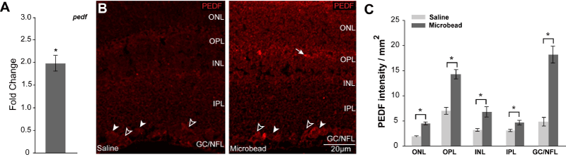
| Figure 3: Elevated IOP increases PEDF expression in murine retina. A. Graphical representation of changes in pedf total mRNA in retina from saline- and microbeadinjected mice, as measured by qPCR. Y-axis represents the fold-change in Δ-CT values for pedf (normalized to the control gene gapdh) microbead-injected, as compared to saline-injected. B. Representative micrographs of immunolabeling for PEDF in longitudinal sections of retina from saline- (right panel) and microbeadinjected (left panel) mice demonstrate increased intensity of labeling in all layers of retina from microbead-injected eyes, as compared to saline-injected eyes. The pattern of PEDF immunolabeling is consistent with RGC soma in the GCL (white arrowheads) and Müller cell processes and endfeet in the ONL and NFL (black arrowheads), respectively. C. Retinal layer-specific quantification of PEDF labeling, expressed in intensity (arbitrary units) per area (mm2). All asterisks denote p<0.05. ONL: Outer Nuclear Layer; OPL: Outer Plexiform Layer; INL: Inner Nuclear Layer; IPL: Inner Plexiform Layer; GCL: Ganglion Cell Layer; NFL: Nerve Fiber Layer |