|
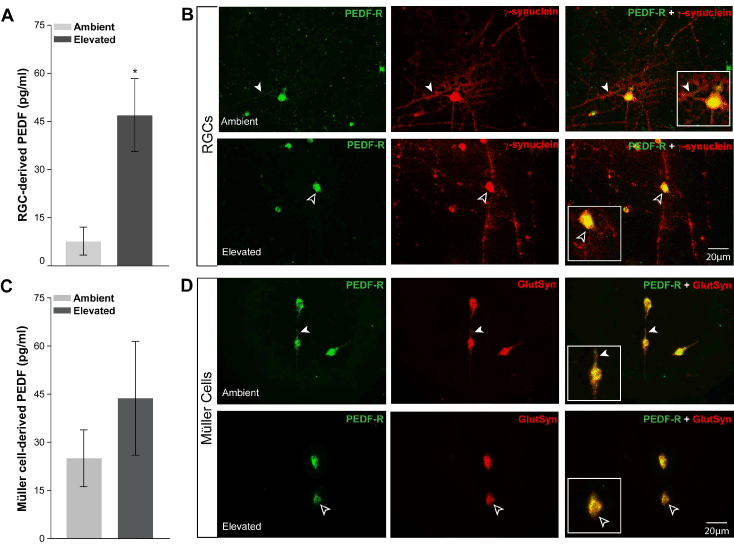
| Figure 4: Elevated pressure alters PEDF production and localization in RGCs and Müller cells in vitro. A. Graphical representation of PEDF concentration (pg/ml; y-axis) in media from primary cultures of purified RGCs exposed to ambient or elevated pressure for 48 hours, as measured by multiplex ELISA. Asterisk denotes p<0.05. B. Fluorescent micrographs of PEDF (green) and γ-synuclein (red) immunolabeling in purified, primary cultures of RGCs exposed to either ambient (top panels) or elevated (bottom panels) pressure for 48 hours reveals a reduction in localization of PEDF to neurites following exposure to elevated pressure (black arrowheads) versus ambient pressure (white arrowheads). C. Graphical representation of PEDF concentration (pg/ml; y-axis) in media from primary cultures of purified Müller cells exposed to ambient or elevated pressure for 48 hours, as measured by multiplex ELISA. D. Fluorescent micrographs of PEDF (red) and glutamine synthetase (green) immunolabeling in purified, primary cultures of Müller cells exposed to either ambient (top panels) or elevated (bottom panels) pressure for 48 hours reveals retraction in cellular processes that is associated with a reduction in PEDF staining following exposure to elevated pressure (black arrowheads), as compared to ambient pressure (white arrowheads). |