 |
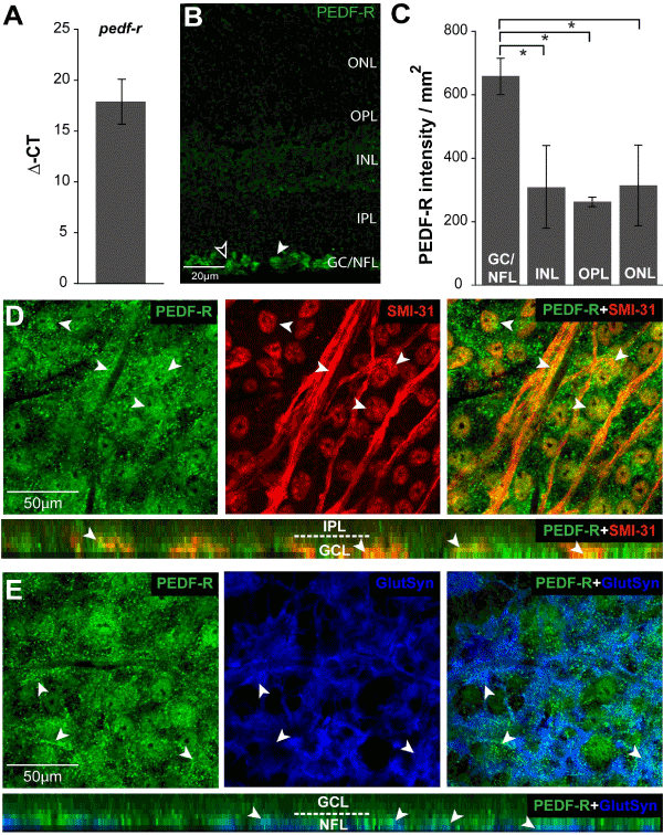
| Figure 5: PEDF-R is constitutively expressed by RGCs and Müller cells in healthy retina. A. Graphical representation of pedf-r total mRNA levels in naïve C57 retina as measured by qRT-PCR. Y-axis represents the average delta of threshold cycle (Δ-CT) values for pedf-r normalized to the control gene gapdh. B. Representative micrograph of longitudinal sections of retina from naïve mice immunolabeled with antibody against PEDF-R. Morphology consistent with localization to RGCs (white arrowheads) and Müller cells (black arrowheads). C. Retinal layer-specific quantification of PEDF-R labeling, expressed in intensity (arbitrary units) per area (mm2). All asterisks denote p<0.05. D. Representative confocal micrographs with orthogonal view (bottom panel) of wholemount retina from naïve mice co-immunolabeled with antibodies against PEDF-R (green) and the RGC marker SMI-31 (red). PEDF-R localizes to SMI-31+RGC soma and axons, as indicated by the yellow appearance of punctate immunolabeling (arrowheads). E. Representative confocal micrographs with orthogonal view (bottom panel) of wholemount retina from naïve mice coimmunolabeled with antibodies against PEDF-R (green) and the Müller cell marker glutamine synthetase (GluSyn; blue). PEDF-R co-localizes with Müller cell endfeet in the NFL, as indicated by the white appearance of immunolabeling (arrowheads). ONL: Outer Nuclear Layer; OPL: Outer Plexiform Layer; INL: Inner Nuclear Layer; IPL: Inner Plexiform Layer; GCL: Ganglion Cell Layer; NFL: Nerve Fiber Layer. |