|
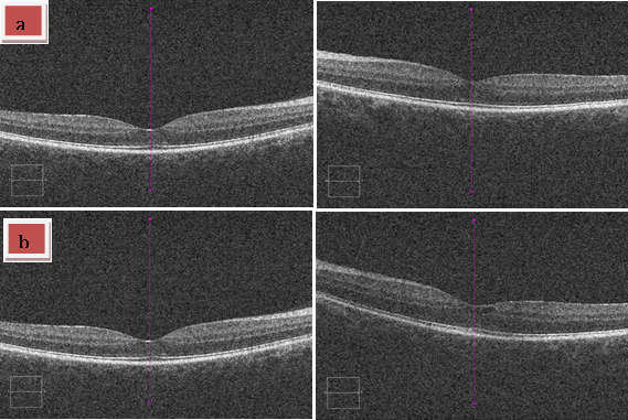
| Figure 4: Macular OCT was normal after treatment in case 1. Patient reported in case 2 condition improved regarding the presence of serous detachments/subretinal fluid although a few intraretinal peri-foveal cysts still remained. a) Patient 1. b) Patient 2. |