|
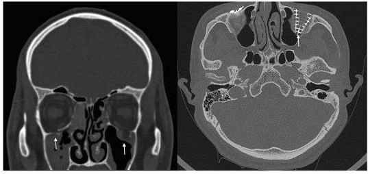
| Figure 3: CT scan orbit in coronal view in patient 5 showing fracture floor of orbit both sides with incarceration of inferior rectus in coronal view both sides (white arrows, left panel). Right panel shows post-operative CT orbit, axial view with intact Titanium mesh, better visible on the left side (white arrow). |