|
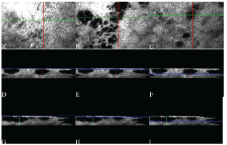
| Figure 5: 3-D analysis at superficial (A) and deep layer (B) (red line) of medically treated eyes, showing smaller (12 μm) microcysts with respect to surgically treated eyes in Y (C, D green line) and X scans (E, F, blue line). The red line identifies the scan plane with respect to the surface (top image). |