|
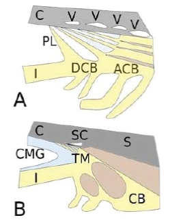
| Figure 3: Enhanced central vision coincides with newly acquired accommodation capabilities. Engrossment of the ciliary muscle impedes the access of aqueous to the posterior angular plexus and modifies the chamber structures in a way that partially relates facility of outflow with ciliary muscle activity. This dependency may impact outflow in the age of presbyopia. A Chamber angle in low mammals with detached and attached portions. A variable number of vessels in the angular plexus evacuates the aqueous humor, after permeating a portion of connective tissue with cells and coloidosmotic GAGs in the extracellular material. B Chamber angle in hominids with uveo-scleral and corneo-scleral meshwork, but not detached ciliary body. The angular plexus has been substituted by a single anterior vessel. The ciliary muscle pulls the scleral spur to open the meshwork, a function that diminishes with age. Coloidosmotic GAGs form the cameral mucous gel (CMG) and are left to the uveo-scleral meshwork, with no remaining of the angular connective tissue. C Cornea; V Vessel from the angular plexus, PL Pectinate ligament; I Iris; DCB Detached ciliary body; ACB Attached ciliary body; SC Schlemm’s Canal; S Sclera; CMG Cameral mucous gel; TM Trabecular meshwork; CB Ciliary body (and ciliary muscle). |