 |
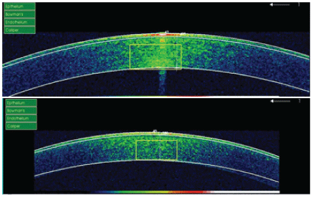
| Figure 3: Corneal OCT of a patient in this study. Visualization of the eye after the performance of corneal OCT. The different layers of the cornea are shown in the upper image. An increased corneal thickness is shown a after application of the artificial tear in the image below. Thus, hydration depends mainly on the corneal epithelium, although a visible increase of the other layers is also observed. |