|
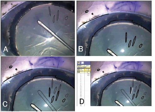
| Figure 2: Measurement of lens movement distance using the overlay method. The images show weakness in the form of a 180° dehiscence. Still images were captured when the anterior capsule was grasped (A) and when the first tear was created (B); then, the cortical opacity, needle, pupil contour, and conjunctival pigmentation were each traced using dotted (A) and solid (B) lines. The dotted lines from (A) were superimposed onto (B), and proper positioning was adjusted by the pupil and pigmentation. The difference between the dotted and solid lines represents the lens movement distance (C). The needle thickness (red line) was matched at 0.4 mm to measure the movement distance (white line) (D). |