|
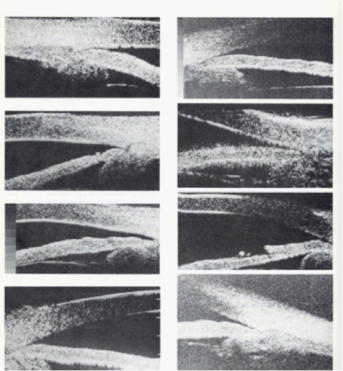
| Figure 2: Ultrasound biomicroscopic images of typical angle cross sections in eyes with the palateau iris syndrome. These images show variations in degree of angle closure and in configuration of the iris and ciliary processes. They all have in common an anterior positioning of the ciliary processes, which produces an absent ciliary sulcusand consequent narrowing of the angle. |