|
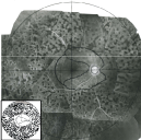
| Figures 5: Panoramic FFA of the right eye of Patient 2 in group II with a long duration before PRP. PRP was performed according to the protocol, but actually the density was 36.0% (inset). IOP temporally decreased from 34 mmHg to 18 mmHg, but increased again to 25 mmHg. IOP did not return to the normal level despite additional PRP to more than 40%, which was achieved 18 months after the diagnosis of NVG. Note the persistence of optic disc neovascularization, which did not disappear, even after the accomplishment of PRP. The missing area of the panoramic photographs (painted grey) in Figure 5 inset is 4.16%. |