 |
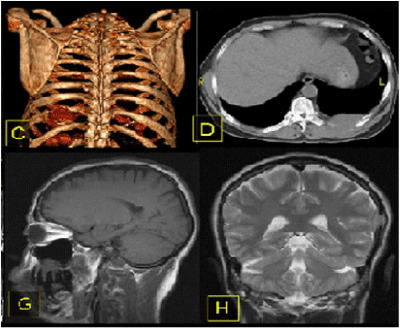
| Figure 3: Extraorbital bony lesions. C) Volume rendered multi-detector CT image of the thorax, seen from the back, and D) transverse CT image reveals a focal osseous lesion of the left tenth rib. G) Sagittal T1-weighted MR image shows a lesion at the soft palate resting on the roots of the upper teeth. H) Coronal T2-weighted MR image of the head shows a focal lesion of the left side of the skull. The lesion has the same signal characteristics as that of the left orbital mass. |