 |
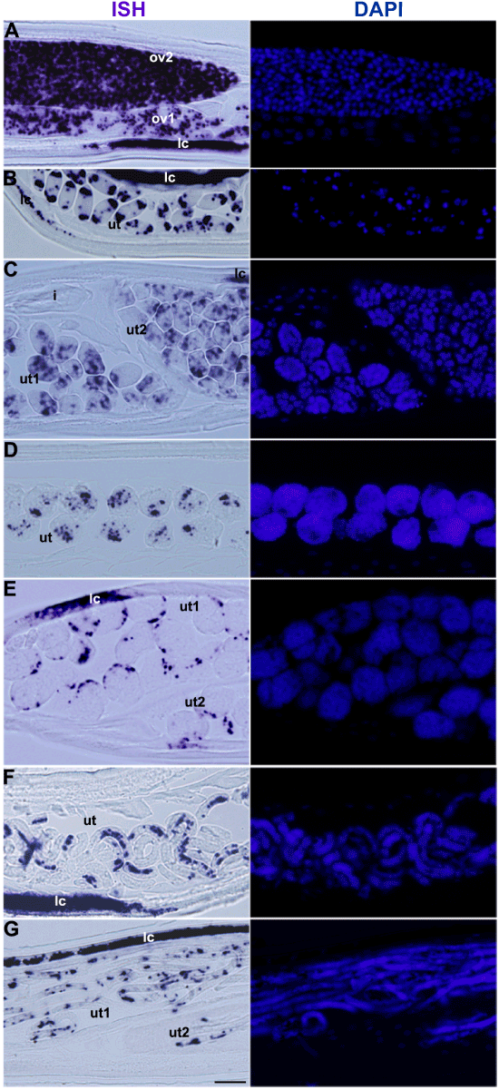
| Figure 6: Wolbachia are asymmetrically segregated into different cells during early embryogenesis and are eliminated from all cells except hypodermal cord cells during morphogenesis. Wolbachia were labeled by ISH using a 16S rRNA probe (left panel), and DNA was stained with DAPI (blue, right panel). Developing oocytes in ov1 are more advanced than in ov2 and embryos in ut1 are more advanced than in ut2. (A) Wolbachia are present in most of the developing oocytes in ovaries; (B) Wolbachia moved predominantly to the poles after fertilization; (C) in early morula stage, Wolbachia are asymmetrically segregated in many blastomeres; (D) in the late morula stage, Wolbachia are present in a smaller number of cells; (E) in comma stage embryos, Wolbachia are absent in most cells and concentrated in hypodermal cord cells; (F) in pretzel stage embryos, Wolbachia were only detected in hypodermal cord cells; (G) in stretched microfilariae, Wolbachia were only visible in hypodermal cord cells. Abbreviations: i: intestine; lc: lateral cord; ov: ovary; ut: uterus; scale bar: 20 μm. |