 |
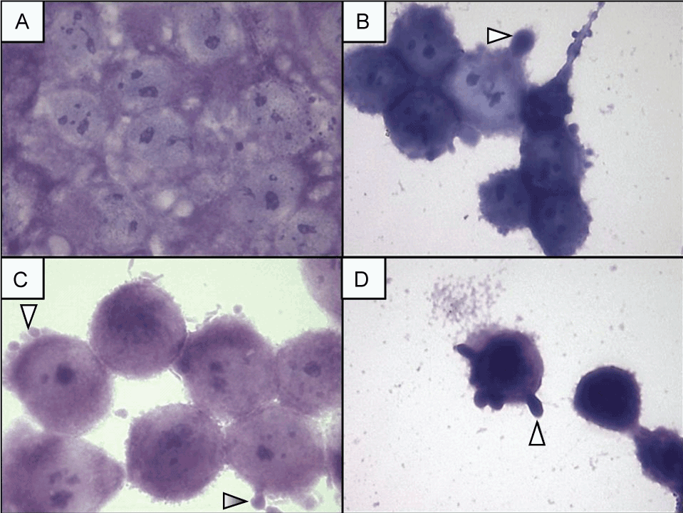
| Figure 2: Adhesion of N.meningitidis in glioblastoma cells line NG97 stained by Giemsa. The line arrows indicate the glioblastoma cells altered by meningococcal adhesion. In (A) the negative control show the NG97 cells line; the adhesion (black arrow) and morphologic alterations, as cytoplasmatic projections (white arrows), caused in (B) by the strain P2143, in (C) by the strain IAL 2443 and in (D) by Y USA. All these meningococci adhesions showed morphologic alterations with blebís formation (white arrows). Amplification of 1000X |