|
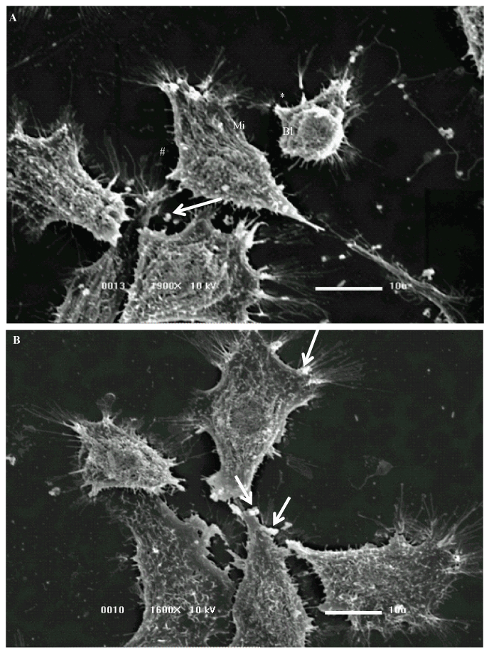
| Figure 3:Scanning electron micrograph of NG97 cell line co-cultured with N. meningitidis (Type B). (A) The NG97 culture were represented by two morphologic distinct cellular types, a small rounded cells and fibroblastic-like (* and #) The cells presented surface alterations like a diminished microvilli (Mi) and blebs (Bl) on the membrane surface. Note the diplococcus (A,B) interacting with the NG97 cell membrane (arrows). |