|
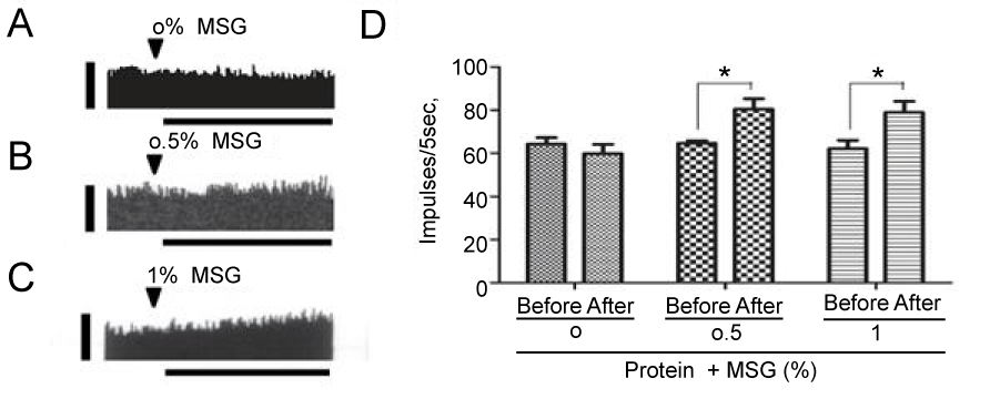
| Figure 3: Vagal gastric activity following intra-gastric application of a protein-rich liquid diet, with or without free glutamate supplementation. (A)- (C): Representative recording of gastric afferent discharge, displayed as a sequential rate histogram, after intragastric administration of 2 mL of a liquid protein-rich (casein) diet supplemented with 0, 0.5, or 1% MSG. Arrowheads indicate the points at which the solution was infused. The vertical bar indicates 100 impulses/5 s. The horizontal bar indicates 30 min. (D): Summary of changes in discharge rate, showing the impulse values of diets containing 0, 0.5, and 1% MSG on gastric afferent activity measured before and after administration. Each point and vertical bar represents means ± SE from 5 different rats * p<0.05 (by a paired t-test). Reproduced from Somekawa et al. [36]. |