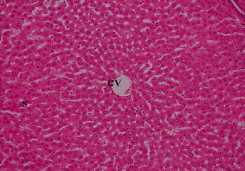
Figure 1:
Section of liver of control animal showing normal structure of hepatic lobule, central vein (cv) and blood sinusoids (s) (HX&E x200).