 |
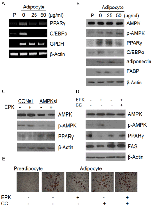
| Figure 2: Effect of EPK on adipogenesis in 3T3-L1 preadipocytes. 3T3-L1 preadipocytes were incubated in medium containing insulin (1.0μg/mL) with or without the indicated concentrations of EPK or CC (compound C). (A) Total RNA was extracted from 3T3-L1cells treated with EPK and used for RT-PCR analysis of PPAγ, C/EBPα, GPDH and β-actin. (B) Total proteins prepared from EPK-treated 3T3-L1 cells were subjected to western blot analysis of AMPK, p-AMPK, PPAγ, C/EBPα, adiponectin, FABP and β-actin. (C) 3T3- L1 cells were transfected with AMPK-siRNA for 48h and treated with or without EPK (50μg/ml) for 24h and were subjected to western blot analysis of AMPK, p-AMPK, PPAγ, β-actin (D) Total proteins prepared from EPK (50 μg/ml) or CC (2 μM) treated 3T3-L1 cells for 6 h were subjected to western blot analysis of AMPK, p-AMPK, PPARγ, FAS and β-actin. (E) Cells were fixed and stained with Oil-Red-O. The Oil-Red-O stained adipocytes were photographed at a ×100 magnification under a microscope. |