|
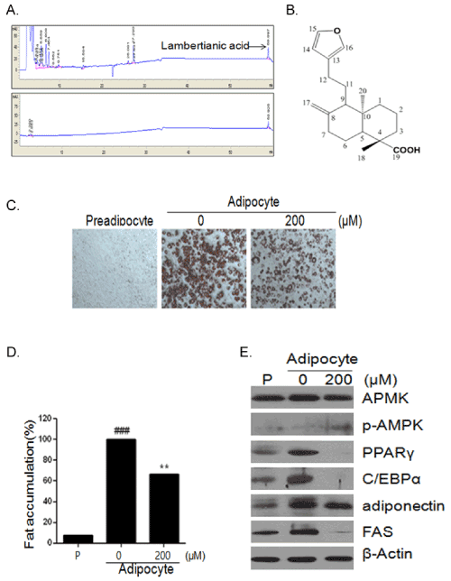
| Figure 5: Effects of LA on adipogenesis in 3T3-L1 preadipocytes. (A) The HPLC analysis of LA in EPK. (B) Chemical structure of LA. (C) 3T3-L1 preadipocytes were treated with various concentrations of LA during differentiation. On day 8, the differentiated cells were stained with Oil-red-O dye and visualized under inverted microscopy at x100 magnifications. (D) After dissolving and cellular lipid retained Oil-Red-O in isopropanol, adipocyte expression was estimated by measuring O.D. using microplate reader (Sunrise, TECAN, Mannedorf, Switzerland) at 510nm. (E) Total protein prepared from LA treated 3T3-L1 (preadipocytes or adipocyte) cells were subjected to western blot analysis of AMPK, p-AMPK, PPARγ, C/EBPα, adiponectin, FABP and β-actin. |