 |
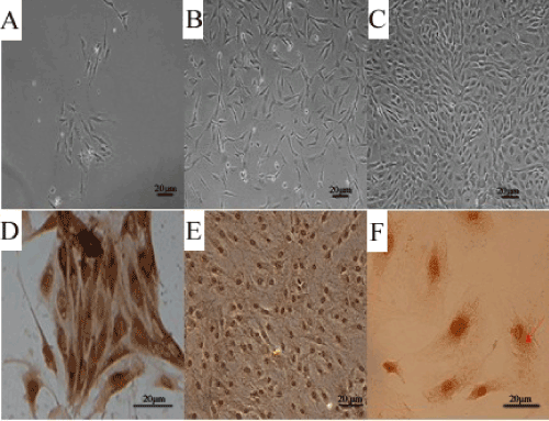
| Figure 1: The primary culture of rat aortic vascular endothelial cells (VECs) and Immunocytochemical staining. Panel A shows 3 days later, a small amount of cells are observable (×100). Panel B shows 6 days later, after replaceing medium ECM, cells are growing rapidly( ×100). Panel C shows 9 days later, when the cells reached 90% confluence it shows that VECs grew with the characteristic ‘cobblestone morphology’ (×100). Panel D, E and F shows these cells were immunostained with factor VIII (D, F×400) antibodies, which is marked antigen of endothelial cells. It demonstrates positive reaction with cytoplasmic brown pigment. No obvious negative cells were seen (E×200). And the factor VIII related antigens were presented as brown granules in the cytoplasm as the red arrow points. D: primary cells; E and F: passage 2 cells. |