|
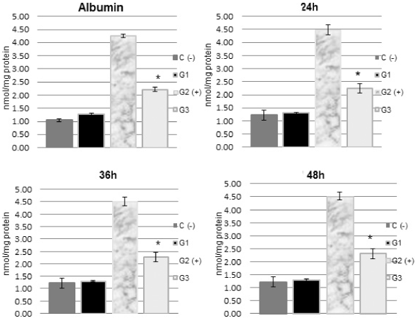
| Figure 4: In vitro treatment of human skin samples with pA decreased UVB-induced MDA. Skin samples were exposed thirty minutes to UVB with or without pretreatment with pA (12%). Non-UVB-exposed skin “n-UVB-”(negative control, C); non- UVB- exposed skin plus pA “n-UVB+”(Group G1); UVB-exposed skin “UVB- “(positive control, Group G2); and UVB-exposed skin plus pA “UVB+” (Group G3). Results are expressed as nmol MDA/mg protein. Data are presented as the means ± SD from six independent experiments. * p<0.001. |