 |
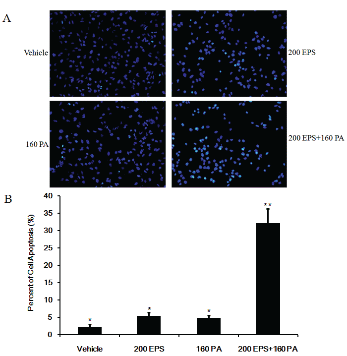
| Figure 3: RBE cells were seeded in a 6-well plate and were treated with 200 μM EPS, 160 μM PA and a combination of 200 μM EPS and 160 μM PA for 24 hrs. Cells were stained with Hoechst and were photographed under a fluorescence microscope (A). The rates of cell apoptosis were calculated based on at least three randomly selected images (B) (*, P<0.05 versus **). |