|
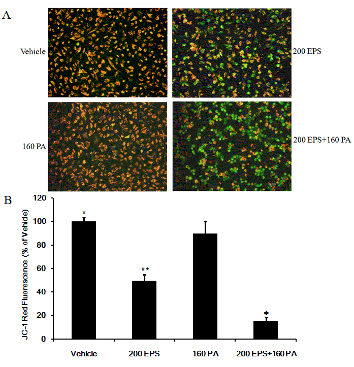
| Figure 5: RBE cells treated with 200 μM EPS, 160 μM PA or a combination of 200 μM EPS and 160 μM PA for 24 hrs were stained with JC-1 and then images (A) were acquired using a Nikon Te2000 microscope (magnification: 200×). (B) The fluorescence intensity was quantified and percent reduction in JC-1 red fluorescence relative to vehicle control cells was calculated. Data from at least 3 randomly selected images are expressed as mean ± SD (*, P<0.05 versus **; +, P<0.05 versus **). |