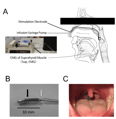
B: Enlarged picture of the electrode tip (filled arrow) connected to a flexible stainlesssteel coil spring tube for electrical stimulation and tip of the polyethylene tube for stimulation by solution (open arrow).
C: Position of the electrode on the posterior wall of the oropharynx and the area stimulated by a test solution. The electrode is indicated by dashed line, and the area after application of a test solution (0.1 ml) is stained blue with food coloring.