|
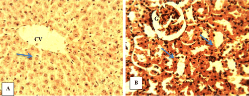
| Figure 5: Photomicrograph of sections of organs from WR-PSGF. (A) Normal hepatic tissue with central vein (CV) and Kupffer cells along the sinusoids (blue arrow). (B) Renal tissue showing normal glomerulus (G) and renal tubules (blue arrows); H&E ×400. |