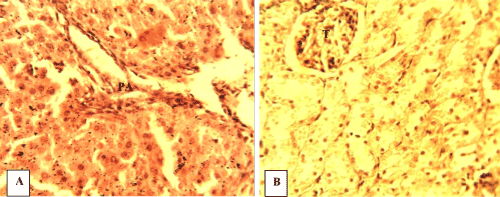
Figure 7:
Photomicrograph of sections of organs from WR-OT. (A) Hepatic tissue showing the portal area (PA) with remarkable histologic change. (B) Renal tissue with moderate degenerated tubules and glomerular tufts (T); H&E ×400.