|
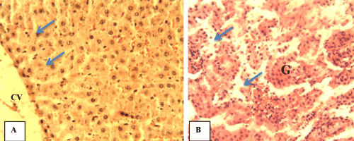
| Figure 9: Photomicrograph of sections of organs from WR-MO. (A) Hepatic tissue showing the central vein (CV) and normal plates of hepatocytes (blue arrows), (B) Renal tissue showing moderate congestion of the glomerulus (G) and interstitium (blue arrows); H&E ×400. |