|
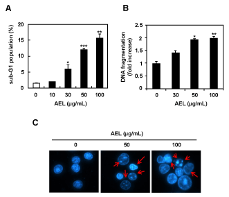
| Figure 2: AEL-induced apoptotic cell death in MCF-7 cells. (A) MCF-7 cells were cultured with different concentrations of AEL for 48 h, fixed and stained with PI, and then the Sub-G1 population was analyzed by flow cytometry. (B) DNA fragmentation was assessed by ELISA. Data values are expressed as mean ± SD of triplicate determinations. Significant differences were compared with the control at *p<0.05, **p<0.01, and ***p<0.001 by Student’s t-tests. (C) Representative images of nuclear condensation in response to AEL treatment as detected by Hoechst staining assay. |