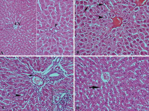
Figure 1: A photomicrograph of sections of liver tissue (A) for control rat shows the normal structure of liver tissue. (B) For a diabetic rat received 50mg Coctus shows congestion of main blood vessels, vacuolar degeneration of hepatocytes (arrowhead) and dilatation of blood sinusoids (arrow). (C) For a diabetic ratreceived 100mg Coctus shows thickening of blood vessels’ wall (arrow), mild
vacuolar degeneration of hepatocytes (arrowhead) and cellular infiltration (in the lower right corner of figure). (D) For a diabetic rat received150mg Coctus shows restoration of normal structure of liver tissue but with mild dilatation of blood sinusoids. (Hx. & E. X 200). |