|
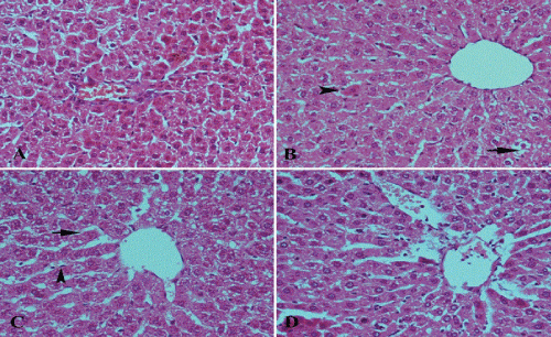
| Figure 2: A photomicrograph of sections of liver tissue (A) for a diabetic rat received (Nano 50) shows distortion of normal architecture of liver tissue, congestion of main blood vessels, dilatation of blood sinusoids and apoptotic and vacuolar degenerated cells. (B) For a diabetic rat received (Nano 100) shows restoration of normal architecture of tissue but with vacuolar degeneration of some cells (arrow) and acidified cells (arrowhead). (C) For a diabetic rat received (Nano 150) shows dilatation of blood sinusoids (arrow) with increased number of Kupffer cells (arrowhead). Minimal degree of vacuolar degeneration is observed. (D) For a diabetic rat receivec insulin shows marked dilatation and congestion of blood sinusoids with no signs of vacuolar degeneration. (Hx. & E. X 200). |