received 100mg Coctus shows mild amelioration of islands of Langerhan. (D) For a diabetic rat received 150mg Coctus shows restoration of normal size and shape with no evidence of vacuolar degeneration. (Hx. & E. X 200).
 |
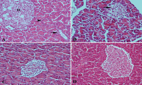
| Figure 3: A photomicrograph of sections of pancreatic tissue (A) for a control rat shows islands of Langerhan (IL), serous acini (arrowhead) and ducts (arrow). (B) For a diabetic rat received 50mg Coctus shows atrophy of islands of Langerhan (arrow) with vacuolar degeneration of many of its cells. (C) For a diabetic rat received 100mg Coctus shows mild amelioration of islands of Langerhan. (D) For a diabetic rat received 150mg Coctus shows restoration of normal size and shape with no evidence of vacuolar degeneration. (Hx. & E. X 200). |