|
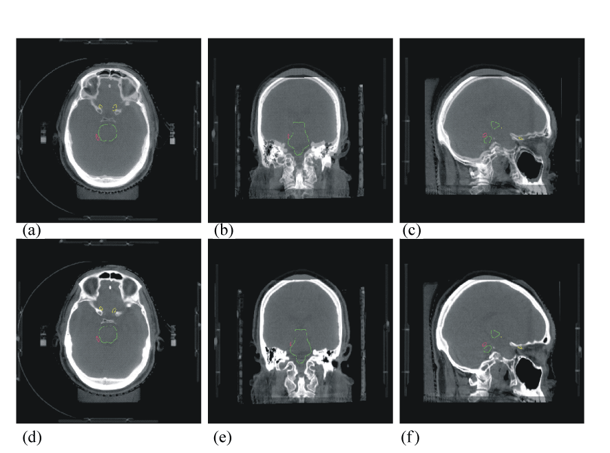
| Figure 8: One example of 6-DOF image-guidance for SRS treatment for Arterio- Venous Malformation (AVM): Top images (a-c) are fused images between planning CT and on-board CBCT after initial setup with BrainLAB mask system and TAPO. The images show the AVM lesion in pink and considerable setup displacements. Bottom images (d-f) show perfect match after the 6D setup correction. |