|
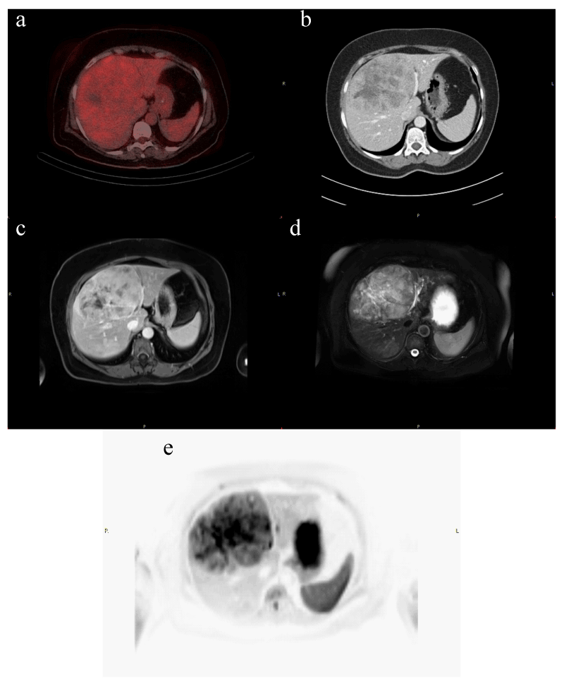
| Figure 1: Registered axial images of patient with a large HCC lesion in the right liver lobe: FDG PET-CT (a), portal phase CECT (b), fat suppression Gd enhanced T1w MRI (c), fat suppression T2w MRI (d), and DWI b800 MRI (e). Note that FDG PET-CT does not show any tracer accumulation in the tumor lesion (non avidity). MRI provides the best tumor to normal liver contrast. |