 |
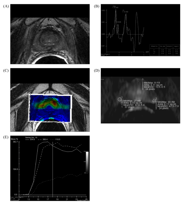
| Figure 1: Positive multi-modality MRI study (patient no. 1). A. Negative endorectal T2-weighted MRI. B. MR spectroscopy showing a suspicious lesion in both lobes (Cho+Cr/Ci ratio = 3). C-E. Dynamic contrast-enhanced MR study showing suspicious lesions in the peripheral zone of the prostate with a typical cancer wash-out slope. |