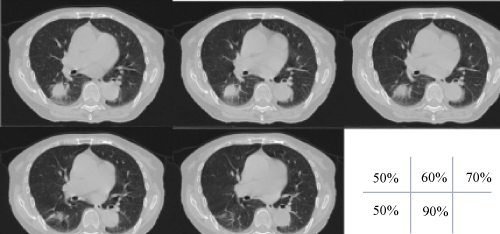
Figure 9:
For the example of a lung tumor, the CT images show each of the respiratory phase images obtained by using 4DCT. The 50%-phase image is defined as the end-exhalation phase image.