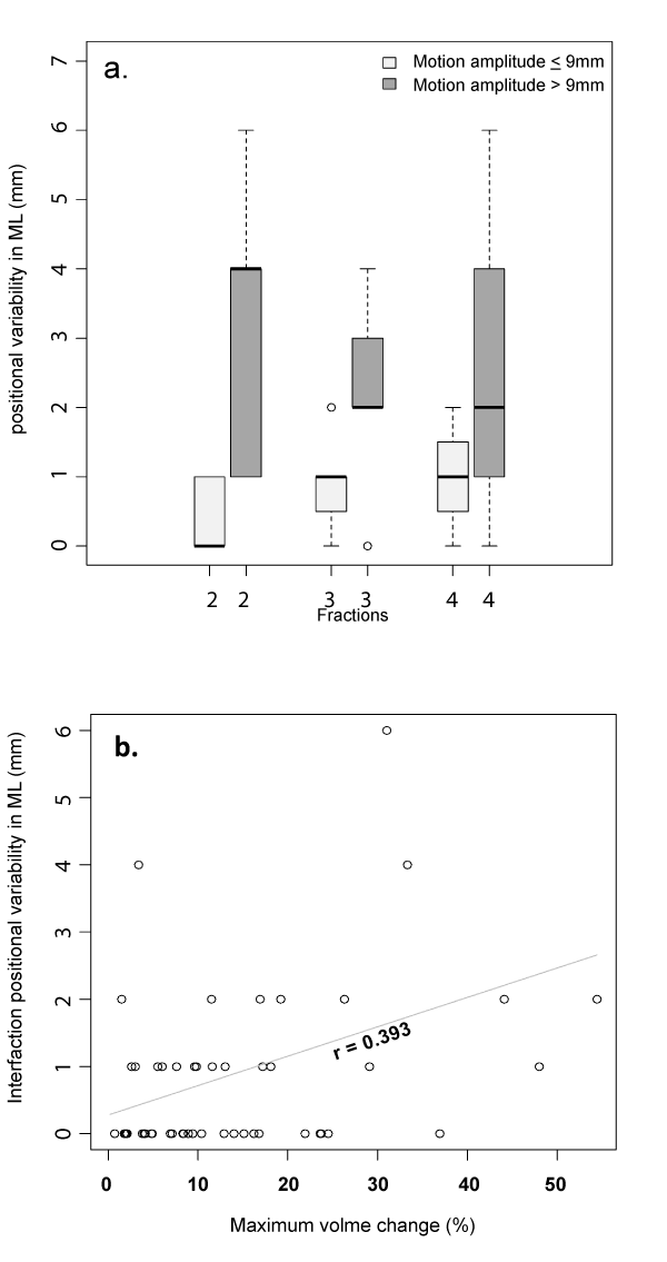
Figure 3:
a and b: Interfraction displacment. Represent increased interfraction displacement in the setting of a) tumor amplitude greater than 9 mm and b) percent volume change per fraction and patient relative to the CBCT of the first fraction.