 |
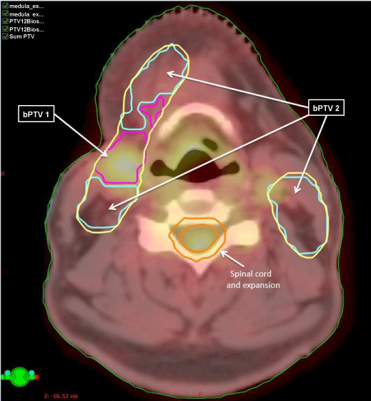
| Figure 4: Fused FDG-PET/CT transversal section of a clinical head and neck cancer patient. Delineated, initial clinical target volume (yellow) and the corresponding biological planning target volumes based on the distribution of FDG-PET/CT (blue and magenta). Organs at risk are spinal cord and expansion (orange). |