 |
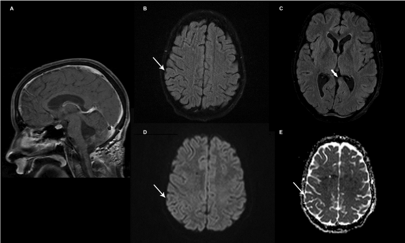
| Figure 2: Images from follow-up MRI study following surgical resection, chemotherapy and after completion of radiation therapy showing changes of surgical resection of posterior fossa mass (A). Axial FLAIR images showing resolution of the right parietal lesion (B-thin white arrow) and decreased conspicuity of the left medial temporal lesion (C-thick white arrow). No evidence of restricted diffusion in the location of the previously seen right parietal lesion on B=1000 diffusion image (D) and ADC map (E) consistent with resolution. |