|
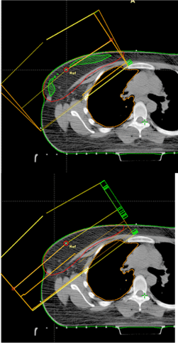
| Figure 2: Showing axial CT simulation images. A: Physical wedge (superior) showing Maximum target dose in green occupying large volume of breast PTV in red. B: Field in field (inferior) resulted in complete disappearance of Maximum target dose from breast PTV. |