|
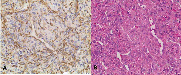
| Figure 2: Pathological manifestations of cranial HPC (x200). A: a staining IHC, vimentin-positive. B: HE staining tumour cells, round or irregular shape, occasionally arranged in turbine-like, nuclear fission or nuclear shaped nuclei is more common, tumour mass is rich in blood vessels, and blood vessel walls thin. Lumen are only coated with a thin layer of the flat endothelial cells, and some abnormal blood vessels characterized with expansion. |