|
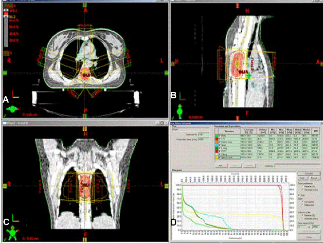
| Figure 4: Typical treatment plan for 3D-CRT for HPC of the upper spine lesion (T4-T8) in (A) axial view, (B) sagittal view, (C) coronal view, and (D) dose-volume histogram (DVH). The patient received postoperatively radiation therapy by 3D-CRT technique consisting of 45Gy in 1.8Gy per fraction in total 25 fractions in continuous irradiation using Linac with 6MV-X rays (Varian 21 EX). The area to be irradiated is determined by correlating preoperative imaging studies, intraoperative findings, and the pathological review of the type. The target volume was generally defined as the tumour bed. For macroscopic residual tumours, the planning target volume (PTV) included the area of contrast enhancement adding a safety margin of 10 mm. The 95% PTV isodose line encompassed the target volume. |