|
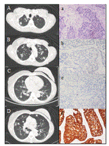
| Figure 1: CT of chest and history of pulmonary nodules. CT of the chest showed multiple bilateral pulmonary metastases. A to D, axial views from apex to basal region of the lung. Fine needle aspiration of the lung nodules showed moderately differentiated adenocarcinoma on H&E staining (a). Immunohistochemical staining of the lung nodule biopsies for: (b), estrogen receptor, (c), progesterone receptor, and (d), positive for high estrogen responders (HER)2/neu. Original magnification, x400. |