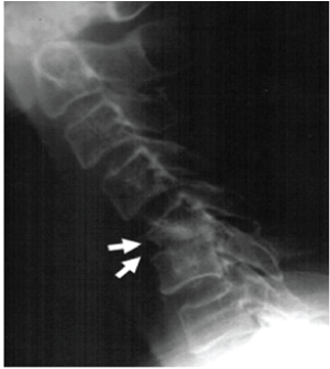
Figure 2:
Magnetic resonance imaging (MRI) showed low signal density on T1-weighted images, indicating vertebral collapse with cord compression at the level of C5. Arrows in the figures indicate the location of damage.