 |
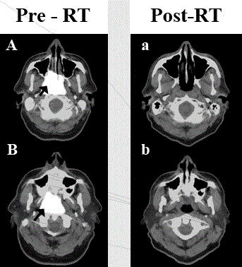
| Figure 1: A representative patient treated with RT with NHL of nasopharynx imaged with [18F] Fluorodeoxyglucose (FDG)– Positron Emission Tomography (PET). A and B are two representative slices pre-RT treatment while a and b are the corresponding slices post-RT. The mass was FDG avid at SUV of 22.2 (black arrow) and corresponded to a soft tissue mass on CT (Figure 2). The patient had complete resolution by 10 months with no increased SUV on the PET. |