|
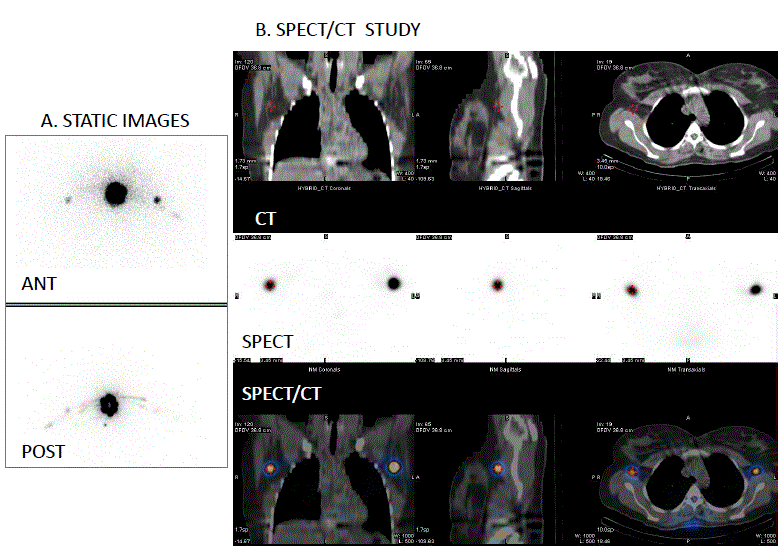
| Figure 1: Sentinel lymph node mapping of a patient with malignant melanoma of the posterior upper trunk (right back). Static images (A., anterior and posterior) and B. coronal CT, SPECT and fused SPECT/CT images obtained after intradermal injection of 99mTc radiocolloid around the melanoma lesion. Static images reveal two drainage pathways of the melanoma lesion and two positive nodes. On SPECT/CT images it is clear that there are two sentinel lymph nodes, one in the right axilla and the other in the left axilla (Nuclear Medicine Department of Patras University Hospital). |