|
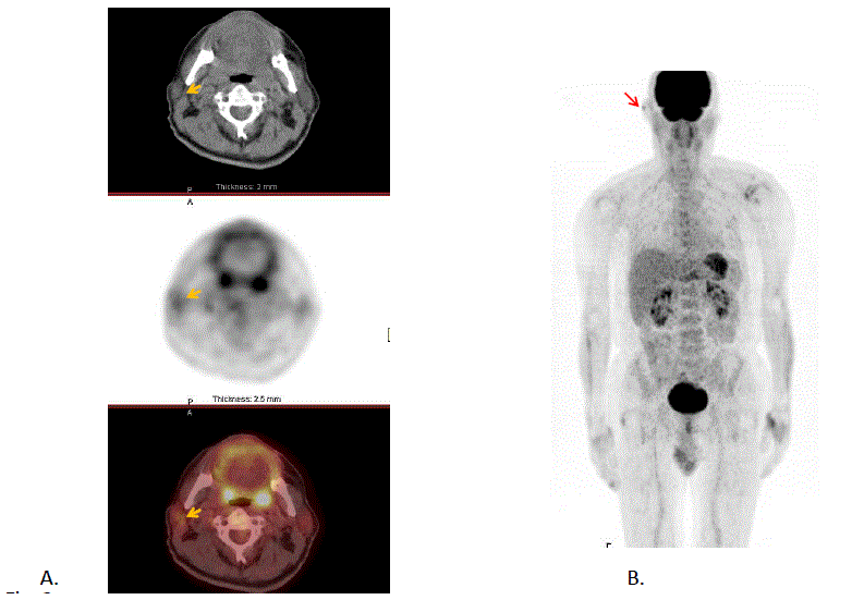
| Figure 2: Malignant Melanoma of the right external ear (surgically excised 2 months prior to PET/CT scan) with positive SLNB (stage IIIA). Initial staging. FDG PET/CT study: axial sections of CT, PET and PET/CT (A), as well as Maximum Intensity Projection (MIP) (B) of the attenuation corrected PET study are shown. A focus of FDG tracer uptake is present in the right ear (red arrow) (SUVmax=2.1) due to postoperative inflammatory changes at the site of resection. There is a very small focus of tracer uptake corresponding to a right intra-parotid lymph node (yellow arrow) (SUVmax=2.0) . The lymph node was negative on needle biopsy, consistent with a reactive inflammatory lymph node (PET/CT department of the Biomedical Research Foundation of the Academy of Athens). |