 |
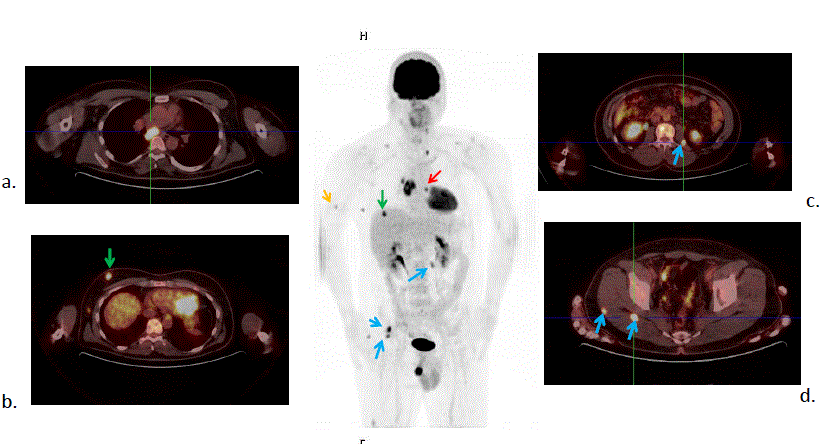
| Figure 4: Patient with malignant melanoma of the left posterior auricular region (stage IIIB), surgically treated 5 months ago. Restaging. FDG PET/CT study: transverse sections of PET/CT, as well as Maximum Intensity Projection (MIP) of the attenuation corrected PET study are shown. Multiple sites of metastasis are noted in cervical lymph nodes, in mediastinal lymph nodes (subcarinal and right hilum) (a.) , in a left lung nodule (red arrow), in soft tissues (c, d, blue arrows) and in multiple cutaneous and subcutaneous nodules of the thoracic wall (b) (one of these nodules, is projecting over the liver on the MIP image, green arrow) and right upper arm (yellow arrow) (PET/CT department of the Biomedical Research Foundation of the Academy of Athens). |