 |
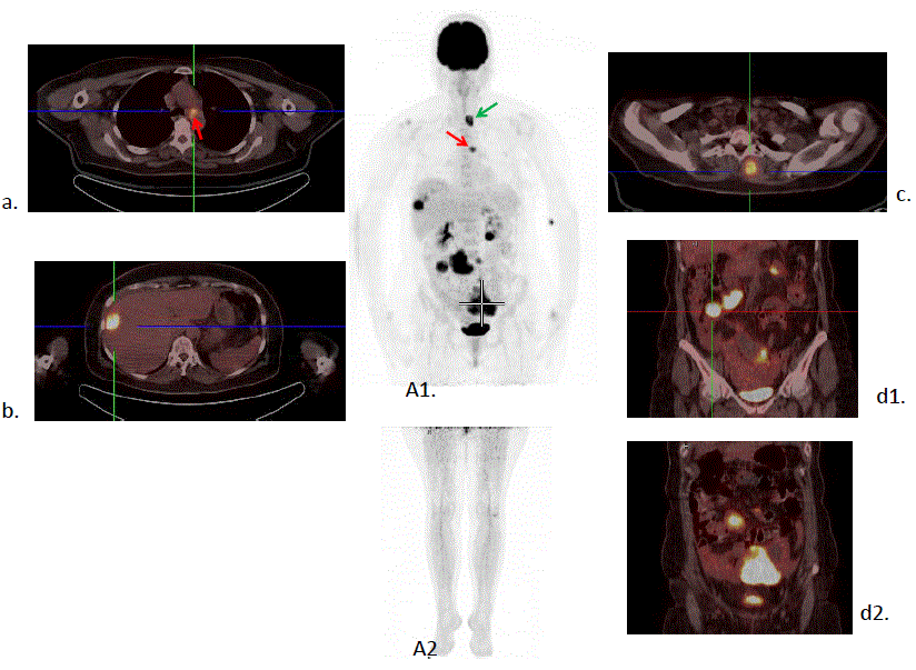
| Figure 5: Patient with malignant melanoma (stage IIIB) of the left upper arm, surgically treated 5 years ago. Metastasis in small bowel, surgically treated 3 years ago. Restaging. FDG PET/CT study: Axial and coronal sections of PET/CT, as well as Maximum Intensity Projection (MIP) of whole body (A1) and feet (A2) are shown. Multiple sites of metastases are noted in a mediastinal lymph node (a., A/P window, red arrow), in the liver (b), in the abdomen (peritoneum and small bowel involvement) and in rhomboideus major muscle (c., green arrow) (PET/CT department of the Biomedical Research Foundation of the Academy of Athens). |